|
|
| |
 |
CADHERIN
SCHEMATIC
|
|
|
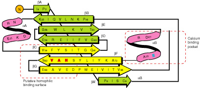 |
| Schematic
drawing of the topology of the first N terminal
cadherin repeat. bA, bB, bE, and bD form a beta
sheet and are shown in green; bC, bF, and bG form
another beta sheet and are shown in yellow; the
alpha helices are shown in magenta. Red type indicates
involvement in adhesion; blue type, calcium binding.
The putative homophilic specificity and calcium
binding pocket are enclosed in dotted lines. |
|
|
|
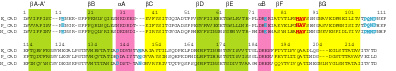
| Alignment
of the first two N terminal cadherin repeats.
|
|
| |
| |
|
|
| |
|
|