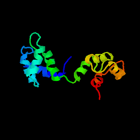
PDB ID: 1DMO
Medline Link
PDB file
Calmodulin-binding Target Database contains known targets of calmodulin.
Biology
Intracellular Calcium ions
Intracellular Ca2+ serves as a common secondary messenger in response to neuronal or hormonal stimuli. Once a cell receives a specific stimulus, the intracellular Ca2+ concentration increases. The increase in Ca2+ concentration initiates many physiological processes.Calmodulin
Calmodulin (CaM) is a common mediator of Ca2+ signals. It is present in almost all types of cells. When intracellular Ca2+ concentration increases, calmodulin binds Ca2+, changes its own conformation, and interacts with one of many targets. This binding activates many enzymes, including phosphodiesterases which are involved in cAMP-singalling pathway, myosin light chain kinases which are used in regulation of motility, and numerous other targets such as nitric oxide synthases, calmodulin-dependent kinases, and calcineurins.Structure
Calcium-free calmodulin (apo-CaM) is composed of four EF-hand motifs (EF-1 through 4 from the N terminus). EF-hand motif is a helix-loop-helix structural motif which is capable of binding Calcium ions in the loop region. EF-3 and EF-4 in the C-terminal domain have higher affinity for Ca2+ ions. Binding of Ca2+ on one of the two EF-hand motifs in each domain facilitates the second binding of Ca2+ by the other EF-hand motif (Ca2+ binds cooperatively in each domain).There are two key features in this structure. First, the relative orientation of helices in each EF-hand motifs are parallel. Second, the linker region between the two domains is very flexible. Because of the flexible linker region, each domain essentially tumbles independently of each other.
In this structure (apo-CaM), there are three hydrophobic patches on the surface. On the C-terminal domain, there are two hydrophobic patches. First one is comprised of Met124, Ala128, Val136 and Met144. There is no matching hydrophobic patch on the N-terminal domain. The second one is found on the N-terminal domain, and is comprised of Ile9, Phe16, Phe65, Pro66, Leu69 and Ala73. The corresponding face of the C-terminal domain contains a rather scattered hydrophobic residues comprised of Ala102, Ala103, Ile125 and Ile130.
The N-terminal hydrophobic patch is partially surrounded by acidic residues such as Glu6, Glu7, Glu14, Asp64, and Glu67. The major hydrophobic patch of the C-terminal domain is also surrounded by acidic residues, Glu119, Glu120, Glu123, Glu127, Glu139 and Glu140. These hydrophobic but negatively charged surface is thought to be responsible for apo-CaM's ability to interact with several IQ-motif containing targets such as neuromodulins and brush border myosin I.
CaM does not have the ability to activate its physiological target proteins
without calcium ions because EF-hand motifs are not in a proper conformation.
Structural studies of Ca2+-bound CaM reveal that upon binding
calcium ions, EF-hand changes its conformation (helices become perpenticular
to each other), and the resulting change in conformation leads to a formation
of deep hydrophobic pockets through which CaM recognizes its physiologic
targets.